Nuclear Medicine
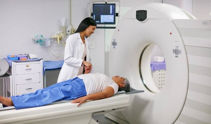
Nuclear medicine is a medical specialty involving the
application of radioactive substances in the diagnosis and treatment of
disease. Nuclear medicine imaging, in a sense, is "radiology done inside
out" or "endoradiology" because it records radiation emitting
from within the body rather than radiation that is generated by external
sources like X-rays. In addition, nuclear medicine scans differ from radiology,
as the emphasis is not on imaging anatomy, but on the function. For such
reason, it is called a physiological imaging modality. Single photon emission
computed tomography (SPECT) and positron emission tomography (PET) scans are
the two most common imaging modalities in nuclear medicine.
In nuclear medicine imaging, radiopharmaceuticals are taken
internally, for example, intravenously or orally. Then, external detectors
(gamma cameras) capture and form images from the radiation emitted by the
radiopharmaceuticals. This process is unlike a diagnostic X-ray, where external
radiation is passed through the body to form an image.
Nuclear medicine tests differ from most other imaging
modalities in that diagnostic tests primarily show the physiological function
of the system being investigated as opposed to traditional anatomical imaging
such as CT or MRI. Nuclear medicine imaging studies are generally more organ-,
tissue- or disease-specific (e.g.: lungs scan, heart scan, bone scan, brain
scan, tumor, infection, Parkinson etc.) than those in conventional radiology
imaging, which focus on a particular section of the body (e.g.: chest X-ray,
abdomen/pelvis CT scan, head CT scan, etc.). In addition, there are nuclear
medicine studies that allow imaging of the whole body based on certain cellular
receptors or functions. Examples are whole body PET scans or PET/CT scans,
gallium scans, indium white blood cell scans, MIBG and octreotide scans.
- Radiopharmacy and Radiochemistry
- Physics of Nuclear Medicine Instrumentation
- Nuclear Medicine Techniques and Special Procedures
- Quality Assurances in Nuclear Medicine
- Recent advances in Nuclear Medicine Techniques
- Radiation Biology
- Radiation Safety in Nuclear Medicine
Recent Published
Submit Manuscript
To give your manuscript the best chance of publication, follow these policies and formatting guidelines.


